Ultrasound Technology
Definition and Basics
Ultrasound Tech innovation, otherwise called sonography, is a clinical imaging strategy that utilizes high-recurrence sound waves to create pictures of the body. It is broadly utilized for indicative purposes since it is harmless, effortless, and doesn’t include ionizing radiation. Ultrasound Tech is frequently utilized to envision muscles, ligaments, veins, and inside organs, assisting with diagnosing different circumstances and guiding specific operations.
History of Ultrasound Tech
The starting points of Ultrasound Tech innovation can be traced back to the mid-twentieth century, with huge commitments from the field of submerged sonar innovation created during The Second Great War. The disclosure of the piezoelectric impact by Jacques and Pierre Curie in 1880 was essential for the improvement of ultrasound. The primary clinical uses of ultrasound showed up during the 1940s and 1950s, reforming the field of symptomatic imaging by permitting clinicians to see inside the body without a medical procedure.
Key Parts of Ultrasound Tech
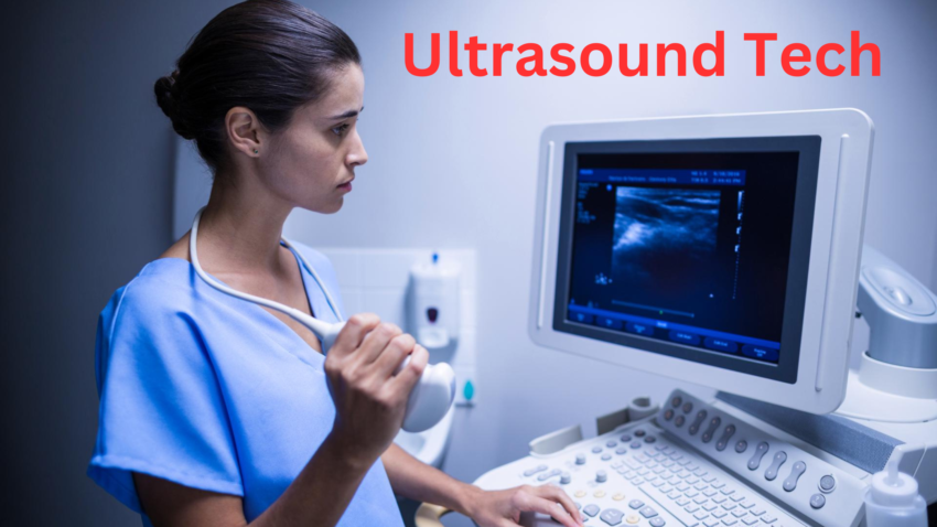
An Ultrasound Tech machine comprises a few key parts, including a transducer, a PC, and a presentation screen. The transducer is the gadget that sends and gets sound waves; it changes electrical energy into sound waves, which travel through the body and bounce off tissues. The returning reverberations are changed over once again into electrical signs, which the PC cycles to make continuous pictures shown on the screen. The quality and clearness of these pictures rely upon the recurrence of the sound waves and the complexity of the handling programming. Also, please visit my other post, Electronic Health Record Systems.
How Does Ultrasound Tech Function?
Ultrasound technology functions by using high-frequency sound waves to create images of the inside of the body. A device called a transducer emits these sound waves, which bounce off tissues and organs. The returning echoes are captured by the transducer and converted into electrical signals, which are then processed by a computer to produce real-time images on a screen.
Standards of Sound Waves
Ultrasound Tech time depends on the ideas of sound waves, specifically the way that they travel through unmistakable media and bounce off surfaces. The transducer emanates high-recurrence sound waves that infiltrate the edge and jump off tissues and organs. These pondered waves, or repeats, are then caught by the transducer. The time it takes for the reverberations to return is estimated and used to choose the space of the reflecting surface from the transducer, shaping the reason for the imaging system.
Picture Development Interaction
The strategy of framing an ultrasound photo incorporates a few stages. To start with, the transducer conveys sound waves, which venture through the casing and mirror again after experiencing tissues and organs. The returning reverberations are caught through the transducer and changed over into electric cautions. These pointers are then handled by utilizing the ultrasound gadget’s PC, which computes the distance and force of the reverberations to make a noticeable representation of the inward frameworks. This real-time picture is shown in a presentation, considering prompt translation and assessment.
Types of Ultrasound Imaging
Ultrasound imaging includes various systems, every ideal to unmistakable symptomatic wishes. The most widely recognized type is 2D ultrasound, which creates level, – layered photos of the interior designs. Further developed procedures comprise three-dimensional ultrasound, which offers 3-layered photographs providing more significant subtlety, and 4D ultrasound, which gives the component of time to make stay-development depictions. Doppler ultrasound is one more specific shape that actions the float of blood through vessels, offering fundamental information about development and recognizing conditions along with blockages or clumps.
Types of Ultrasound Techniques
There are several types of ultrasound techniques, each serving specific diagnostic purposes. Doppler ultrasound measures blood flow and velocity in vessels, helping detect blockages and other vascular issues. 3D ultrasound creates three-dimensional images of internal structures, providing detailed views of organs and fetal development. 4D ultrasound adds dimension to time, capturing real-time moving images that are often used in prenatal scans.
2D Ultrasound Tech
2D Ultrasound Tech is the most unusual shape of ultrasound imaging, producing flat, -dimensional pix of inner organs and tissues. It is substantially used for recurring diagnostic examinations. 2D ultrasound provides excellent stability between picture quality and accessibility, making it a flexible device in medical diagnostics.
Three-D and 4D Ultrasound Tech
3-D Ultrasound Tech takes the traditional 2D pictures a step further by way of creating 3-dimensional pictures. This approach offers extra specific perspectives of structures, providing intensity and a higher expertise of spatial relationships. 4D Ultrasound Tech adds the detail of time to 3-D imaging, generating stay, actual-time three-D photos. This is particularly beneficial in obstetric examinations to study the actions and expressions of the fetus, supplying a stronger reveal for looking forward to mother and father and greater distinct diagnostic data for healthcare providers.
Doppler Ultrasound Tech
Doppler Ultrasound Tech measures the float of blood via vessels, supplying valuable statistics about blood circulation. It detects the route and velocity of blood glide, which is vital for diagnosing conditions together with blood clots, blocked arteries, and heart valve defects. Doppler ultrasound may be completed by the use of diverse modes, including colouration Doppler, energy Doppler, and spectral Doppler, each imparting exceptional sorts of facts approximately blood float dynamics.
Uses of Ultrasound Innovation in Medication
Ultrasound innovation is widely used in medicine for diagnostic and therapeutic purposes. It allows for real-time imaging of internal organs, aiding in the diagnosis of conditions related to the heart, liver, kidneys, and other vital organs. In obstetrics, ultrasound monitors fetal development and detects any abnormalities during pregnancy.
Analytic Imaging
Ultrasound Tech is extensively utilized for demonstrative imaging in various clinical fields. It works with imagining inside organs, finding anomalies and guide strategies. Normal purposes envelop inspecting the stomach, pelvis, heart, veins, and outer muscle gadget. Ultrasound’s real-time imaging usefulness makes it a significant gadget for surveying organ capability, recognizing cancers, pimples, and various abnormalities, and following the advancement of infections.
Remedial Purposes
Past diagnostics, Ultrasound Tech has a few mending programs. Extreme focus designated ultrasound (HIFU) is utilized to manage growths with the guide of warming and obliterating harmful tissues without the need for obtrusive careful treatment. Moreover, lithotripsy utilizes sound waves to separate kidney stones into more modest pieces that can be passed normally. Remedial ultrasound additionally incorporates physiotherapy programs, where sound waves are utilized to advance tissue rebuilding and lessening contamination in conditions like tendinitis and muscle lines.
Interventional Strategies
Ultrasound Tech is instrumental in directing interventional methods, comprehensive biopsies, needle yearnings, and catheter arrangements. Its continuous imaging usefulness takes into consideration exact zeroed-in, limiting the gamble of difficulties and upgrading the precision of those methodologies. For example, sooner or later a biopsy, or ultrasound can manually the needle to the exact area of the conventional tissue, guaranteeing that the example assembled is illustrative of the district of the subject. Additionally, in vascular mediations, ultrasound can help region catheters and guide wires fittingly, improving the security and viability of the way.
Ultrasound in Obstetrics and Gynecology
In obstetrics and gynaecology, ultrasound is a vital tool for monitoring and assessing female reproductive health. It is commonly used to visualize the fetus during pregnancy, allowing healthcare providers to track fetal growth, detect congenital abnormalities, and monitor the placenta and amniotic fluid levels.
Pre-birth Ultrasound Tech
Pre-birth ultrasound is a basic device for following fetal improvement and wellbeing eventually of being pregnant. It finds inherent peculiarities, examines fetal development, and chooses the newborn child’s capability. Routine sweeps are executed at different levels of being pregnant, comprising of the first-trimester ultrasound to insist on being pregnant and gauge the due date, the second-trimester oddity test to test for formative issues, and the 0.33-trimester ultrasound to survey fetal increment and amniotic liquid degrees.
Gynecological Ultrasound
Gynaecological ultrasound assesses the young lady’s conceptive machine, diagnosing conditions which incorporate fibroids, ovarian sores, and endometriosis. It is utilized to look at the uterus, ovaries, fallopian tubes, and encompassing designs. Transvaginal ultrasound, wherein the transducer is embedded into the vagina, gives more prominent unmistakable photographs of the pelvic organs and is specifically gainful for early pregnancy tests and assessing pelvic throb or odd dying.
Evaluating Fetal Wellbeing
Standard ultrasounds at some stage in pregnancy play a fundamental capability in evaluating fetal well-being. They uncover fetal increment, investigate formative inconveniences, and inspect the levels of amniotic liquid. Doppler ultrasound can assess the blood float inside the umbilical wire and fetal vessels, giving fundamental measurements about the child’s prosperity. These tests assist medical services suppliers with finding capacity cerebral pains early, remembering ideal intercessions and better administration of unnecessary risk pregnancies.
Ultrasound in Cardiology
In cardiology, ultrasound, specifically echocardiography, is a crucial diagnostic tool used to assess the heart’s structure and function. It enables cardiologists to visualize the heart’s chambers, valves, and blood flow, helping detect abnormalities such as heart valve disorders, congenital heart defects, and cardiomyopathies.
Echocardiography
Echocardiography, for the most part, called a reverberation, utilizes ultrasound to make certain photographs of the heart. It assesses coronary heart capability, size, and shape, and distinguishes irregularities. Transthoracic echocardiography (TTE) is the typical strategy, wherein the transducer is situated at the chest. It bears the cost of complete realities about the heart’s chambers, valves, and blood coast designs, aiding the conclusion and the board of different cardiovascular circumstances like cardiovascular breakdown, valvular confusion, and innate coronary heart surrenders.
Stress Echocardiography
Stress echocardiography surveys how the heart’s abilities under pressure, every now and again, are caused by means of exercise or medicine. This investigation empowers findings that may not be seen when the heart is unwinding, comprehensive of coronary vein problems. During the check, depictions are taken when the heart is haggard to look at the distinctions in the heart trademark. This aids in diagnosing ischemia, which happens while blood drift to the heart muscle is diminished, and might manually cure decisions for circumstances at any point like angina and myocardial dead tissue.
Transesophageal Echocardiography (TEE)
Transesophageal echocardiography (TEE) includes placing a specific test into the throat to get exact photos of the coronary heart. This strategy gives more clear perspectives than standard echocardiography, in light of the fact that the throat is found near the coronary heart, decreasing the impedance from ribs and lungs. TEE is particularly helpful for evaluating the aorta, and heart valves, and identifying clusters or masses in the coronary heart. It is often utilized in victims with muddled heart conditions, all through cardiovascular surgery, and in seriously sick victims in which novel imaging is imperative.
Ultrasound in Crisis Medication
In emergency medicine, ultrasound is an indispensable tool for rapid, non-invasive diagnosis and treatment. It allows emergency physicians to quickly assess patients with trauma by detecting internal bleeding, fluid accumulation, and organ damage through a FAST (Focused Assessment with Sonography for Trauma) exam. Ultrasound also diagnoses conditions like pneumothorax, cardiac tamponade, and deep vein thrombosis.
Quick Test
The Engaged Appraisal with Sonography for Injury (Quick) test speedy assesses injury patients for inward dying, especially inside the mid-region and chest. It is a quick, harmless methodology performed at the bedside, making it a priceless device in crisis settings. The Quick test finds ways of life-compromising circumstances alongside hemoperitoneum (blood inside the stomach cavity) and pericardial emission (liquid around the coronary heart), permitting set-off and appropriate intercessions.
Directing Crisis Techniques
Ultrasound distribution crisis strategies include chief line arrangements, thoracentesis (liquid expulsion from the pleural region), and pericardiocentesis (liquid expulsion from the pericardial sac). Its real-time imaging capacity upgrades the precision and assurance of those techniques, diminishing the danger of cerebral pain. For example, an ultrasound-directed focal line position permits clinicians to envision the objective vein, ensuring the right needle inclusion and catheter situation.
Mark of-Care Ultrasound (POCUS)
Mark-of-care ultrasound (POCUS) is utilized by clinicians at the bedside to go with quick findings and treatment decisions, upgrading impacted individual consideration in crisis settings. POCUS can inspect circumstances like pneumothorax (imploded lung), cardiovascular tamponade (resist the heart from liquid development), and stomach aortic aneurysm. Its conveyability and straightforwardness of purpose make it a flexible gadget for crisis doctors, permitting quick evaluation and the executives of essentially sick victims.
Progressions in Ultrasound Innovation
Advancements in ultrasound technology have significantly enhanced its diagnostic and therapeutic capabilities. Innovations such as high-resolution imaging, 3D and 4D ultrasounds, and Doppler techniques provide clearer, more detailed images and real-time motion capture. Portable and handheld ultrasound devices have made the technology more accessible, enabling point-of-care diagnostics in diverse settings, from rural clinics to emergency rooms.
Artificial intelligence and AI Incorporation
Man-made brainpower (computer-based intelligence) and machine dominating are upsetting ultrasound by utilizing improving photograph superior grades, aiding examination, and robotizing routine obligations. Artificial intelligence calculations can improve photo choice, see styles, and proposition genuine time examination, diminishing the responsibility on clinicians and developing symptomatic precision. Machine learning to realize models can be taught to secure specific circumstances, providing determination help and prescient examination.
Versatile and Handheld Gadgets
Progressions in age have achieved the improvement of compact and hand-held ultrasound devices, expanding availability and remembering factor-of-care diagnostics in various settings. These smaller gadgets can be used in remote, ambulances, or even in patients’ homes, getting demonstrative abilities the heading the place of need. Compact ultrasound devices likewise are cost strong, making them an engaging option for help obliged settings.
Three-dimensional Printing and Ultrasound
Joining 3D printing with ultrasound time permits the coming of exact physical styles for careful making arrangements and tutoring. Ultrasound pictures might be changed into three-dimensional models, which can be distributed to offer an unmistakable delineation of the life systems. These designs help specialists envision complex designs, plan careful cycles, and practice procedures. They likewise capability significant scholarly instruments for clinical understudies and students.
Instructive Pathways and Vocations
Educational pathways and careers in ultrasound technology, also known as diagnostic medical sonography, typically begin with an associate’s degree or a bachelor’s degree in sonography or a related health science field. These programs include anatomy, physics, and patient care coursework, as well as hands-on clinical training. Advanced certifications in specialized areas like obstetrics, vascular technology, or cardiac sonography can enhance career prospects.

Turning into an Ultrasound Specialist
Becoming an ultrasound specialist requires finishing an approved program, usually a two-year associate degree or an endorsement program. Coursework incorporates life structures, physiology, material science, and hands-on clinical tutoring. A few packages offer single-man reaches, which might provide better preparation and potential outcomes than specialization.
Affirmation and Permitting
Affirmation from organizations such as the American Library for Symptomatic Clinical Sonography (ARDMS) supplements task possibilities and validity inside the area. Certificate ordinarily involves passing really takes a look at that investigate expertise and capabilities in unambiguous areas of sonography. Proceeding with instruction is expected to stay up with the latest progressions inside the area.
Vocation Possibilities and Specializations
Ultrasound professionals can have some expertise in locales that incorporate obstetrics, cardiology, vascular sonography, and outer muscle sonography. The interest in proficient experts keeps on growing, granting several professional prospects in emergency clinics, centres, demonstrative imaging offices, and confidential practices. Professionals likewise can seek jobs in tutoring, examination, and framework deals.
Future Patterns and Developments
Future trends and developments in ultrasound technology are poised to revolutionize medical diagnostics and treatment. Emerging innovations such as AI-enhanced imaging, which leverages machine learning algorithms for more accurate and efficient diagnoses, are at the forefront. Portable and wearable ultrasound devices are expected to become more widespread, enabling continuous monitoring and remote diagnostics. 3D and 4D imaging advances will offer even more detailed and dynamic views of internal structures.
Upgraded Imaging Procedures
Future progressions in imaging techniques guarantee even additional lucidness and analytic exactness, working on quiet outcomes. Strategies that incorporate elastography, which estimates tissue solidness, and evaluation of more prominent ultrasound, which utilizes microbubbles to improve picture top calibre, are being created and are unobtrusive. These upgrades will broaden the indicative skills of ultrasound and deal with more unambiguous records around tissues and organs.
Ultrasound in Non-Clinical Fields
Past cure, ultrasound uncovers applications in fields like business non-unfavorable looking at, submerged investigation, and material innovation. It is utilized to take a gander at the respectability of materials, find blemishes, and screen underlying well-being. In the submerged investigation, ultrasound permits mapping the ocean bottom and finding objects. These non-clinical bundles exhibit the adaptability and far-reaching effect of ultrasound age.
Worldwide Effect and Openness
Endeavours to make ultrasound period more noteworthy and available worldwide can radically affect medical services in underserved regions, improving analytic abilities and patient consideration. Versatile and lower-valued ultrasound contraptions related to preparing applications for medical services transporters can assist with connecting the space in symptomatic contributions. Telemedicine and distant meetings, upheld by ultrasound, can enhance access to agreeable medical services in remote, helpful asset-bound districts.
FAQ About Ultrasound Tech
Q1: What is ultrasound technology?
A: Ultrasound Tech uses high-frequency sound waves to create images of the inside of the body, commonly used for medical diagnostics.
Q2: How does ultrasound work?
A: Ultrasound works by emitting sound waves into the body, which bounce off tissues and organs. These echoes are captured to create real-time images.
Q3: What are the types of ultrasound?
A: Common types include 2D, 3D, 4D, and Doppler ultrasound, each providing different levels of detail for various diagnostic purposes.
Q4: What is a 3D ultrasound?
A: A 3D ultrasound creates three-dimensional images, offering more detailed views of structures. It is often used in obstetrics for fetal imaging.
Q5: What is Doppler ultrasound used for?
A: Doppler Ultrasound Tech measures blood flow through vessels helping diagnose conditions like blood clots and blocked arteries.