Table of Contents
ToggleMacular Degeneration Test
Macular Degeneration
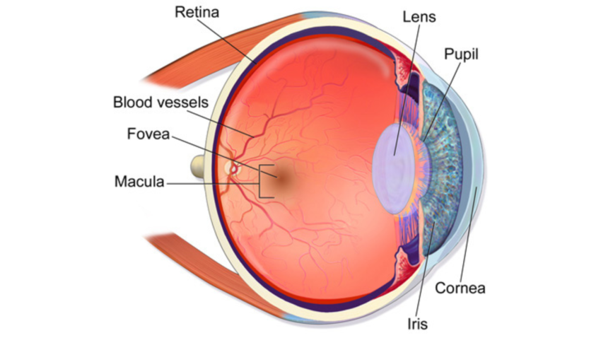
Macular Degeneration Test, also known as age-related macular degeneration (AMD), is a common eye condition that affects the macula, the small central part of the retina. The retina is the layer of tissue at the back of the eye that processes light and sends visual information to the brain. The macula is responsible for central vision, which is crucial for reading, driving, and recognizing faces.
Types of Macular Degeneration
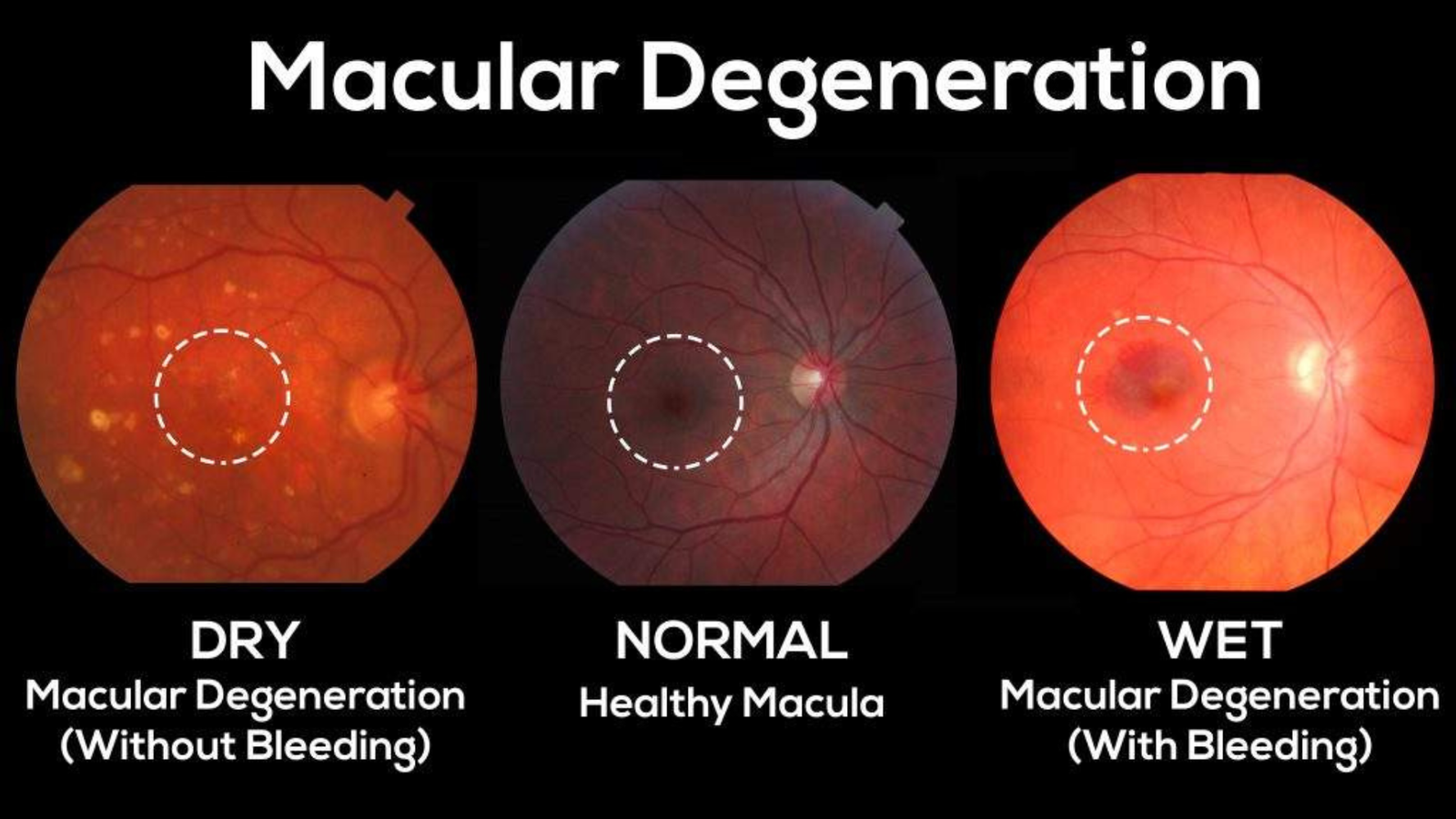
There are two primary sorts of AMD: dry (atrophic) and wet (neovascular or exudative). Dry AMD is more regular and advances gradually. It happens when the macula diminishes over the long run as a feature of the maturing system, prompting a progressive loss of focal vision. Wet AMD is more uncommon but more serious. It occurs when abnormal blood vessels grow under the retina and macula, leading to leakage of blood and fluid, which causes rapid and severe vision loss.
Risk Factors and Symptoms
Several factors can increase the risk of developing a Macular Degeneration Test, including age (50 years and older), family history of AMD, smoking, high blood pressure, high cholesterol, obesity, and prolonged exposure to ultraviolet (UV) light. Common symptoms of the Macular Degeneration Test include blurred or distorted vision, difficulty seeing in low light, and a dark or empty area in the center of vision. Early detection and regular eye examinations are crucial for managing AMD and preserving vision.

Importance of Early Detection
- Timely Intervention: Early detection of macular degeneration allows for prompt treatment, which can slow the progression of the disease and help maintain vision.
- Improved Quality of Life: Identifying the Macular Degeneration Test early helps individuals preserve their ability to perform daily activities, reducing the impact on their independence and overall quality of life.
- Preventive Measures: Early diagnosis provides an opportunity to implement lifestyle changes and preventive strategies to reduce the risk of further vision loss.
Benefits of Early Diagnosis
Early diagnosis of Macular Degeneration Test can significantly impact the management and progression of the disease. Detecting AMD at an early stage allows for timely intervention, which can slow the progression of the disease and help maintain vision. Early treatment options for wet AMD, such as anti-VEGF injections, can prevent further vision loss and, in some cases, improve vision.
Impact on Quality of Life
Macular Degeneration Test can severely impact an individual’s quality of life, challenging everyday tasks and reducing independence. Early detection and appropriate management can help individuals with AMD maintain their quality of life, continue their daily activities, and reduce the risk of severe vision loss.
Preventive Measures
The eyes, from UV light, can help reduce the risk of developing Macular Degeneration Test. Individuals at higher risk should consult their eye care professional for personalized advice on preventive strategies.
Types of Macular Degeneration Tests
- Visual Acuity Test: Measures the sharpness of vision using a Snellen chart to detect changes in central vision.
- Amsler Grid Test: Utilizes a grid pattern to identify distortions or blank spots in the visual field, indicating potential macular damage.
- Optical Coherence Tomography (OCT): Employs light waves to capture detailed cross-sectional images of the retina, revealing early signs of both dry and wet AMD.
Visual Acuity Test
The visual acuity Macular Degeneration Test is a standard eye exam that measures how well a person can see at various distances. It involves reading letters on a chart (Snellen chart) placed at a specific distance. This test helps detect vision changes and is often the first step in diagnosing AMD.
Amsler Grid Test
The Amsler grid Macular Degeneration Test is a simple and quick test used to detect abnormalities in the central visual field. It involves looking at a grid of horizontal and vertical lines to check for distortions, blurriness, or blank spots that can indicate macular damage.
Optical Coherence Tomography (OCT)
Optical coherence tomography (OCT) is an advanced imaging technique that provides detailed cross-sectional images of the retina.OCT helps identify structural changes in the macula and detect early signs of dry and wet AMD. This non-invasive test is critical for monitoring the disease’s progression and evaluating treatments’ effectiveness. Also, visit my other post. Ear Infection Contagious.
Visual Acuity Test
- Purpose and Procedure: The visual acuity Macular Degeneration Test measures the sharpness of vision using a Snellen chart. The patient reads letters from a distance of 20 feet, one eye at a time.
- Interpreting Results: Results are expressed as a fraction, such as 20/20, indicating the testing distance over the distance a person with normal vision can read the same line.
- Common Variations: This includes a near-vision Macular Degeneration Test with smaller charts for reading distance and digital acuity tests using specialized equipment to detect issues not apparent in standard distance testing.
Purpose and Procedure
The visual acuity Macular Degeneration Test measures the sharpness of vision using a Snellen chart, which features rows of letters decreasing in size. The patient covers one eye and reads the letters on the chart from a set distance, typically 20 feet. This process is repeated for the other eye.
Interpreting Results
Results are expressed as a fraction, such as 20/20, where the first number represents the testing distance, and the second indicates the distance a person with normal vision can read the same line. For example, 20/40 vision means that the patient can read at 20 feet, and a person with normal vision can read at 40 feet.
Common Variations
Variations of the visual acuity Macular Degeneration Test include near vision tests using smaller charts held at reading distance and digital acuity tests performed with specialized equipment. These Macular Degeneration Tests help detect vision issues that are not apparent in standard distance testing.
Amsler Grid Test
- How to Use the Amsler Grid: Hold the grid at a reading distance (about 14-16 inches) in good lighting. Focus on the central dot with one eye covered and repeat for the other eye. Note any distortions or missing lines in the grid.
- Signs to Look For: Look for wavy, blurry, or missing lines. Distorted or broken lines or dark spots may indicate macular damage and should be reported to an eye care professional immediately.
- When to Consult a Doctor: If you notice any abnormalities on the Amsler grid, such as lines appearing wavy or parts of the grid missing, consult your eye care professional as soon as possible for a complete assessment.
How to Use the Amsler Grid
The Amsler grid is a simple, hand-held card featuring a grid pattern with a central dot. Patients focus on the central dot and note any areas where the lines appear wavy, broken, or missing. This test can be performed at home or in the clinic and helps identify early signs of macular degeneration.
Signs to Look For
Patients should look for any distortions, such as wavy lines or dark areas, and report these findings to their eye care professional. Such distortions can indicate damage to the macula and require further investigation.
When to Consult a Doctor
If the Amsler grid test reveals any abnormalities, it is crucial to consult an eye care professional immediately. Early detection of changes in the macula can lead to timely treatment and potentially slow the progression of AMD.
Optical Coherence Tomography (OCT)
- Non-Invasive Imaging Technique: OCT uses light waves to capture detailed cross-sectional images of the retina, allowing eye care professionals to see each of the retina’s distinctive layers without the need for invasive procedures.
- Early Detection of Macular Changes: OCT can detect early signs of dry and wet AMD, including thinning of the macula, fluid accumulation, and abnormal blood vessels, making it a crucial tool for early diagnosis and monitoring disease progression.
- Quick and Painless Procedure: The OCT Macular Degeneration Test is quick, typically taking just a few minutes, and is painless, involving no contact with the eye. Patients look at a target while the device scans the retina.
Technology Behind OCT
OCT uses light waves to take cross-sectional retina images, similar to how ultrasound uses sound waves. It provides high-resolution images that allow doctors to see each of the retina’s distinctive layers, enabling early detection of macular changes.
What to Expect During the Test
During an OCT test, the patient rests their chin on a support and looks into the machine at a target. The OCT device scans the eye without touching it, taking a few minutes. The test is painless and non-invasive.
Analyzing OCT Results
Macular Degeneration Test results are analyzed by comparing the patient’s retinal images to normal, healthy retinas. Doctors look for signs of AMD, such as thinning of the macula, fluid accumulation, or abnormal blood vessels. These detailed images help in diagnosing the type and severity of AMD.
Fundus Photography
- Role in Diagnosing AMD: Fundus photography involves capturing detailed images of the retina, macula, and optic nerve, which help document and monitor the progression of macular degeneration over time.
- Procedure and Preparation: The procedure typically requires pupil dilation using eye drops. The patient sits in front of a fundus camera, which takes a series of photographs quickly and non-invasively.
- Understanding Fundus Images: Fundus images reveal key indicators of the Macular Degeneration Test, such as drusen (yellow deposits under the retina), pigment changes, and abnormal blood vessels. They provide a visual record for tracking disease progression and guiding treatment decisions.
Role in Diagnosing AMD
Fundus photography involves taking pictures of the eye’s interior surface, including the retina, macula, and optic nerve. These images help document the condition of the retina and monitor changes over time.
Procedure and Preparation
The procedure requires pupil dilation using eye drops to obtain a clear retina view. The patient sits in front of the fundus camera, and a series of photographs are taken. The process is quick and noninvasive.
Understanding Fundus Images
Fundus images are analyzed for signs of the Macular Degeneration Test, such as drusen (yellow deposits under the retina), pigment changes, and abnormal blood vessels. These images provide a visual record that can be compared over time to track the progression of the disease.
Fluorescein Angiography
- How It Works: Involves injecting a fluorescent dye into the bloodstream to highlight retinal blood vessels.
- Procedure: Pupil dilation, dye injection, and photographing the eye as the dye travels through the vessels.
- Results: Identifies leaking or damaged blood vessels, helping to diagnose and manage wet AMD.

How It Works
Fluorescein angiography involves injecting a fluorescent dye into the bloodstream and photographing it as it travels through the blood vessels in the retina. This test helps identify leaking blood vessels characteristic of wet AMD.
Preparation and Procedure
Patients should fast for a few hours before the macular degeneration test. The procedure starts with pupil dilation and dye injection into a vein, usually in the arm. A special camera photographs as the dye circulates through the retinal blood vessels.
Interpreting Angiography Results
The images captured during fluorescein angiography highlight abnormalities in the retinal blood vessels. Leaking or damaged vessels appear as bright spots, indicating the presence of a wet Macular Degeneration Test and guiding treatment decisions.
Indocyanine Green Angiography (ICG)
- Purpose: ICG visualizes the deeper layers of the retina and choroid, and it is beneficial in detecting occult (hidden) choroidal neovascularization associated with wet Macular Degeneration Test.
- Procedure: The test involves injecting a dye into the bloodstream, which emits infrared light, allowing it to penetrate deeper into the retina. A specialized camera captures detailed images of the retinal and choroidal vessels.
- Benefits: ICG provides high-resolution images of the deeper layers of the eye, identifying abnormal blood vessels that might be missed with other imaging techniques. This makes it valuable in complex AMD cases.
Purpose of ICG
ICG visualizes the deeper layers of the retina and choroid, which are not visible with fluorescein angiography. This Macular Degeneration Test is beneficial in detecting occult (hidden) choroidal neovascularization associated with wet AMD.
Steps Involved in the Test
Like fluorescein angiography, the Macular Degeneration Test involves injecting a dye into the bloodstream. The dye used in ICG emits light in the infrared spectrum, allowing it to penetrate deeper into the retina. A specialized camera captures images of the dye’s passage through the retinal and choroidal vessels.
Benefits of ICG in AMD Diagnosis
The Macular Degeneration Test ICG provides detailed images of the deeper layers of the eye, helping to identify abnormal blood vessels that might be missed with other tests. It is precious in complex cases where other imaging techniques are inconclusive.
Managing Macular Degeneration
- Treatment Options: Anti-VEGF injections, laser therapy, and photodynamic therapy are available for wet AMD, while nutritional supplements and lifestyle changes help manage dry AMD.
- Lifestyle Adjustments: Quit smoking, maintain a healthy diet rich in leafy greens and fish, protect your eyes from UV light, and manage chronic conditions like hypertension.
- Regular Monitoring and Follow-Up: Schedule frequent eye exams to monitor disease progression, adjust treatments, and implement new interventions as needed.
Treatment Options
Treatment choices for AMD rely upon the kind and seriousness of the illness. Dry AMD treatment focuses on slowing progression through lifestyle changes and nutritional supplements. Wet AMD may require anti-VEGF injections, laser therapy, or photodynamic therapy to control abnormal blood vessel growth and leakage.
Lifestyle Adjustments
Lifestyle adjustments, such as quitting smoking, maintaining a healthy diet rich in leafy greens and fish, protecting eyes from UV light, and managing underlying health conditions like hypertension, can help manage AMD and reduce the risk of progression.
Regular Monitoring and Follow-Up
Regular eye exams and monitoring are essential for individuals with AMD. Follow-up visits allow eye care professionals to track the disease’s progression, adjust treatment plans, and implement new interventions as needed.
Conclusion
In conclusion, early detection and management of macular degeneration are essential for preserving vision and maintaining quality of life. Regular eye exams, prompt treatment, and lifestyle adjustments can significantly slow the progression of the disease and reduce the impact on daily activities. By staying informed about the types of tests available and actively participating in preventive measures, individuals at risk of AMD can take proactive steps to safeguard their vision.
FAQs About Macular Degeneration Test
Q1: What are the early signs of macular degeneration?
A: Blurred vision, difficulty seeing in low light, and dark spots in the center of vision.
Q2: How often should I get tested for macular degeneration?
A: Annually if you are over 50 or have risk factors; more frequently if diagnosed with AMD.
Q3: Is there a cure for macular degeneration?
A: There is no cure, but treatments can slow progression and manage symptoms.
Q4: Can lifestyle changes help prevent AMD?
A: Yes, quitting smoking, eating a healthy diet, and protecting the eyes from UV light can reduce risk.
Q5: What should I do if I notice changes in my vision?
A: Contact your eye care professional immediately for a comprehensive eye exam.